Who are the experts. А а B - Cc D E e 10 Which drawing in Figure 41 is streptococci.

Arrangement Of Cocci Shape Bacteria Microbiology Medical Laboratory Science Microbiology Study
View the full answer.
. These are a diverse group of commensal species commonly found orally including S. Which drawing in Figure 41 is a tetrad. We review their content and use your feedback to keep the quality high.
This allows them to adhere. 71Which drawing in Figure 41 is streptococci. Streptococci illustration figure drawing diagram image.
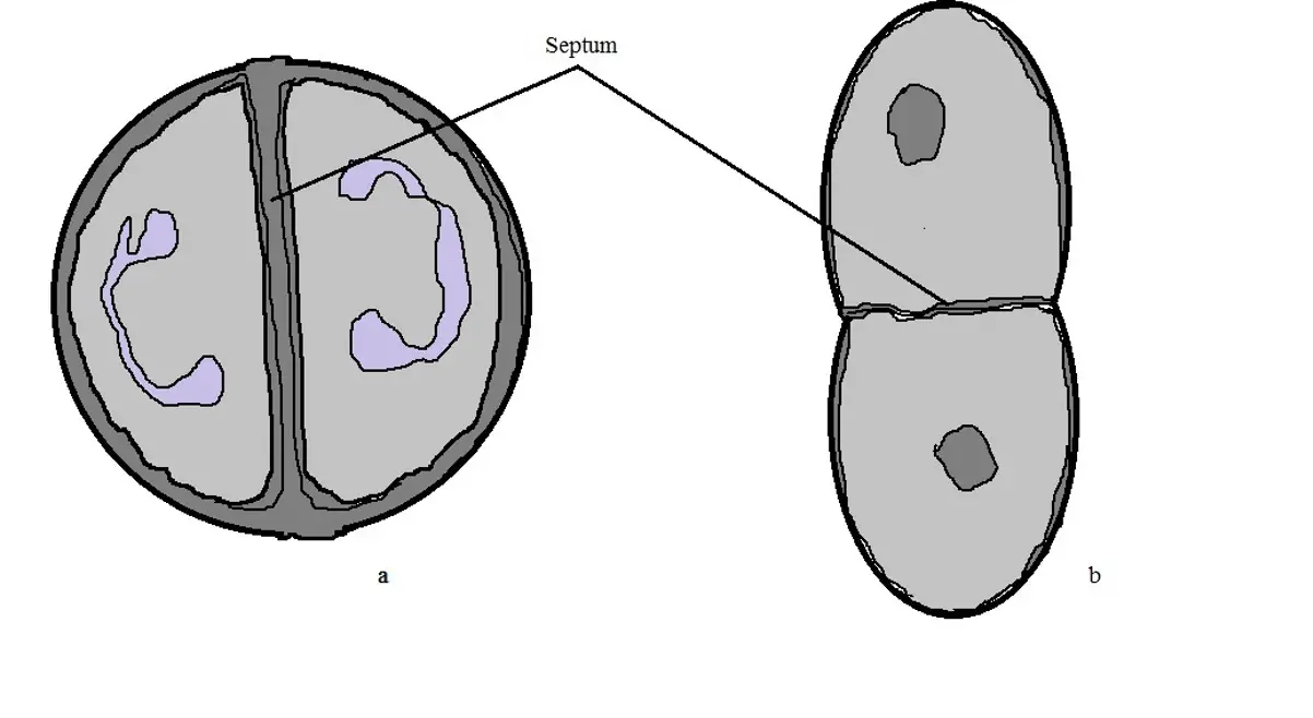
A Their DNA is not enclosed within a membrane. Some strains may produce mucoid colonies Figure 1. Th Drawing by molekuul 1 57 Streptococcus Drawing by Troscha 0 15 Halitosis bad breath.
Which drawing in Figure 41 possesses an axial filament. D strip of cocci shaped bacteria. Clipart by PATTER 2 72 streptococcus bacteria line icon vector illustration Stock Illustrations by vectorwin 0.
Which drawing in Figure 41 is a bacillus. This illustration is included in the following Illustration Toolkit. More than 50 types of S pyogenes M proteins have been identified on the basis of antigenic specificity.
In the figure above which drawing. Both the M proteins and lipoteichoic. PowerPoint Win Mac compatible.
In group A streptococci the R and T proteins may serve as epidemiologic markers but the M proteins are clearly virulence factors associated with resistance to phagocytosis. When incubated aerobically this group of streptococci may render less obvious β-hemolysis also known as α-prime hemolysis which is. Perhaps the most common and logical way to begin a figure drawing is to work from top to bottom - to initially indicate and place the models head.
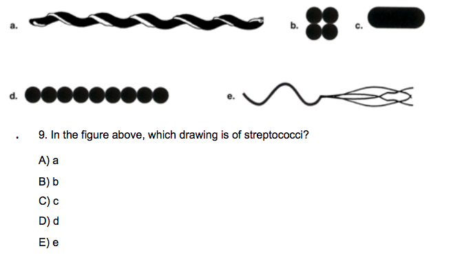
In the figure above which drawing is of streptococci. However once the pneumococcal culture ages 24-48 hours the colonies become flattened and the central portion becomes depressed which does not occur with viridans streptococci Figure 2. They can synthesize dextrans from glucose.
____ Which of the following statements about prokaryotic cells is generally false. They possess 80S ribosomes. Streptococcus pyogenes are spherical to ovoid microorganisms measuring up to 1 μm in diameter.
The cell wall also consists of several structural proteins Figure 13-2. Which drawing in Figure 41 possesses an axial filament. Which drawing in figure 41 is a bacillus.
Mutans and cause endocarditis after release into the bloodstream from tooth extraction figure 12. Experts are tested by Chegg as specialists in their subject area. Agalactiae colonies can be flat grayish-white or orange mucoid and creamy.
Streptococci illustration figure drawing diagram image. Mutans is responsible for approximately half of all cases of bacterial endocarditis. 100 2 ratings Streptococci are gram positive bacteria which are.
Which drawing in Figure 41 is streptococci. Which drawing in Figure 41 is streptococci. Which Drawing In The Figure Is Streptococci.

Chapter Four Microbiology Flashcards Quizlet
Streptococcus Bacteria Classification Shape Infection Gram Stain

2 1 Sizes Shapes And Arrangements Of Bacteria Biology Libretexts Bacteria Prokaryotic Cell Bacterial Cell Structure
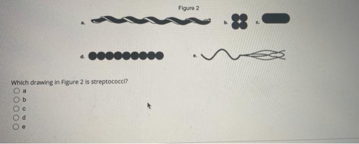
Solved Figure 2 D Which Drawing In Figure 2 Is Chegg Com

Pin On Enregistrements Rapides
Solved A B C D E 9 In The Figure Above Which Drawing Chegg Com

Pin On Zebook2

Streptococcus Bacteria Classification Spherical Shapes Of Bacteria Cocci Mor Sponsored Paid Paid Bacteria Spheric Bacteria Shapes Bacteria Shapes
0 comments
Post a Comment